Mammografi
Lad os give dig klarhed om dit bryst med en klinisk mammografi
Vi hjælper dig med at få et klart svar på, om der er knuder i brystet vha. røntgen, klinisk undersøgelse og ultralydsscanning. Vi har markedets nyeste udstyr, der giver stor præcision og sikkerhed.
Klinisk mammografi
Vi hjælper dig med at få et klart svar på, om der er knuder i brystet vha. røntgen, klinisk undersøgelse og ultralydsscanning. Vi har markedets nyeste udstyr, der giver stor præcision og sikkerhed.
Klinisk mammografi
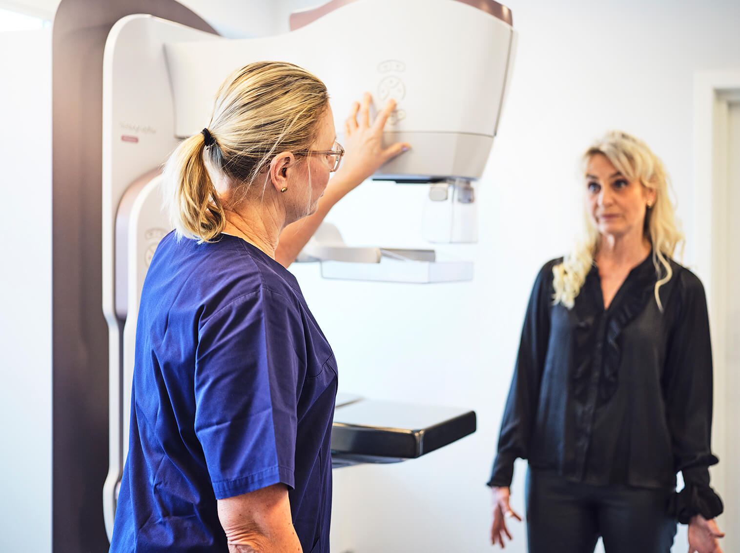
En klinisk mammografi foretages for at se efter knuder i brystet.
Undersøgelsen består af:
Mammografi, som er en røntgenundersøgelse af brysterne.
Klinisk undersøgelse, hvor brysterne undersøges for knuder.
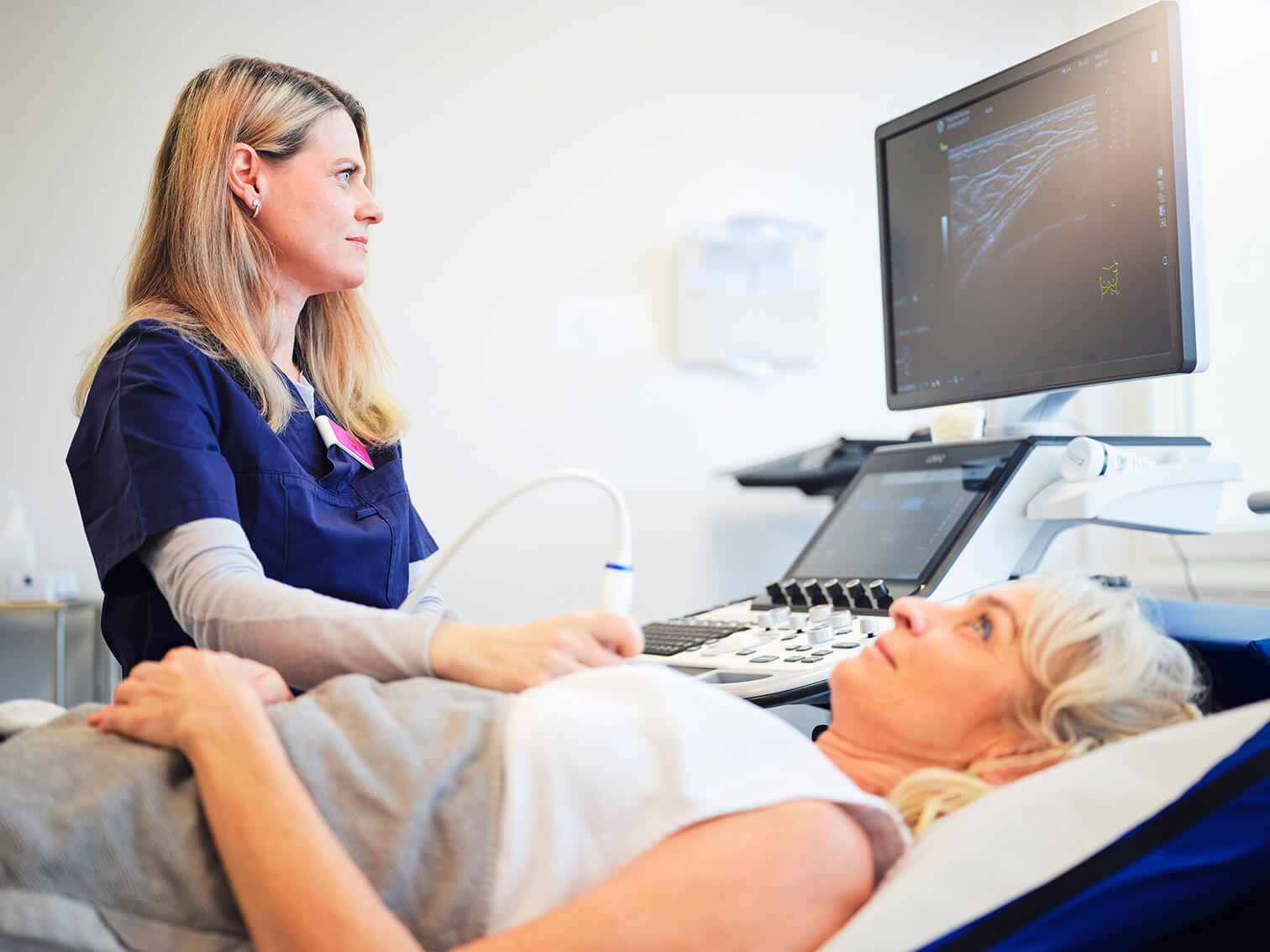
Ultralydsskanning af bryst og armhuler.
Se vores liste med de oftest stillede spørgmål og svar om mammografi her.
Hvornår bør der foretages klinisk mammografi:
• Ved knude/følelig ændring i brystet
• Ved nytilkommen indtrækning af hud eller brystvorte
• Ved spontan blødning eller klar væske fra brystvorten
• Efter betændelse hos ikke-ammende kvinder
• Ved sår på brystvorte, der ikke vil hele
• Som led i kontrol efter operation for brystkræft
• Hos genetisk disponerede
• Ved vedvarende ensidigt hævede lymfeknuder i armhule
• Ved fund i brysterne ved fx CT eller PET-skanning
Selve undersøgelsen:
Ved mammografien bliver brystet kortvarigt presset fast mellem 2 plader, hvorefter der tages røntgenbilleder af hvert bryst.
Efter mammografien undersøger røntgenlægen brysterne for følelige forandringer og foretager en ultralydsskanning af brysterne.
Såfremt røntgenlægen finder en forandring i brystet, bør denne undersøges nærmere ved en vævsprøve (biopsi). Er der behov for at tage en vævsprøve, vil dette blive gjort med det samme.
Varighed
Der skal regnes med ca. 1⁄2-1 times ophold på klinikken.
Undersøgelseskrav
Klinisk mammografi udføres udelukkende på patienter, der er henvist fra læge/hospital.
Undersøgelsessted
Undersøgelsen finder sted hos:
Progardia Mamma
Hedegaardsvej 88 2300 København S
Pris inkl. ultralydsscanning fra 2.550,-
Klinisk mammografi udføres på dig, der er henvist fra læge/hospital
Kontakt Progardia for mere information via vores kontaktformular eller ved at ringe til os på:
+45 70 60 11 60.
Vores leverandører:


Hvor længe varer undersøgelsen?
Man skal regne med ca. ½ time til mammografi og ultralyd. Såfremt der tages biopsi/vævsprøve kan undersøgelsen tage længere tid.
Skal jeg forberede mig til undersøgelsen?
Kan man få lavet mammografi, når man har implantater?
Får man svar på undersøgelsen samme dag?
Hvornår tages biopsi/vævsprøve?
Hvornår er der svar på biopsi/vævsprøver?
Må jeg lave fysisk aktivitet efter en biopsi/vævsprøve?
Må jeg have en pårørende med til undersøgelsen?
Er der parkeringspladser i nærheden af klinikken?
Er der metrostation i nærheden af klinikken
Ring eller skriv
Vi sidder klar til at hjælpe dig, hvis du har spørgsmål
Du kan også finde svar på de mest almindelige spørgsmål på siden spørgsmål og svar.